The human body is composed of trillions of cells, and within the nucleus of each cell lies the genetic blueprint that governs our biological functions, traits, and characteristics. This blueprint is contained within the chromosomes, which are tightly packaged structures made up of DNA (deoxyribonucleic acid) and proteins. Understanding the fundamentals of chromosome and packaging of DNA is crucial for comprehending the intricate mechanisms that regulate gene expression, cell division, and heredity.
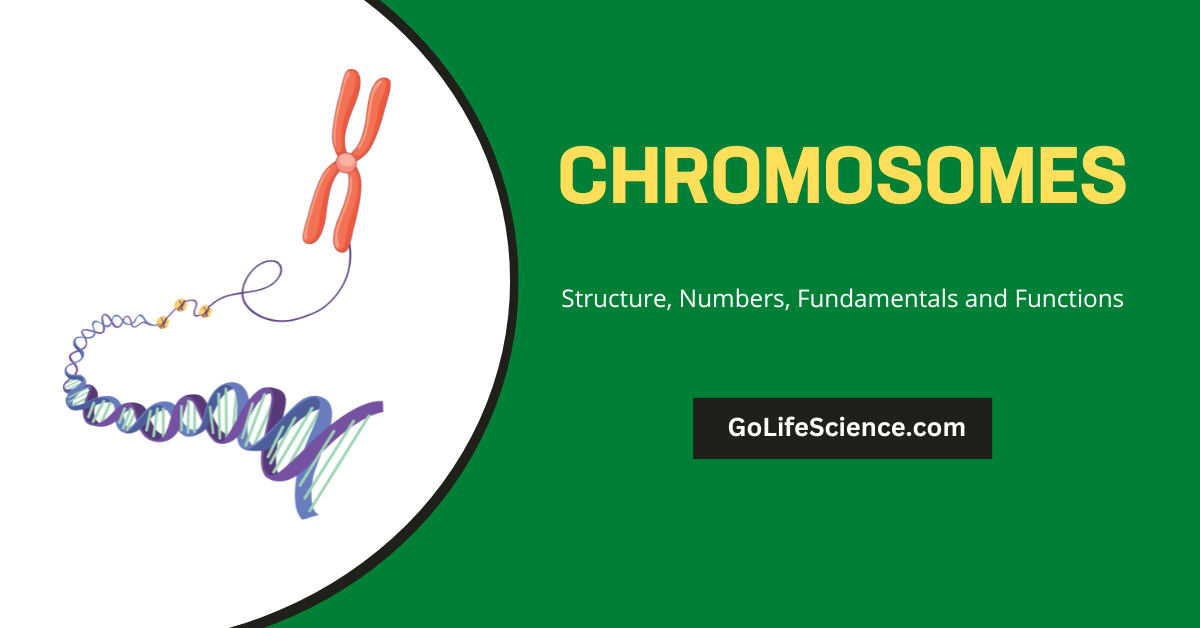
Chromosomes are the fundamental units of genetic information in living organisms. These microscopic structures, composed of DNA and proteins, play a crucial role in heredity, development, and the overall functioning of cells. In this comprehensive article, we will delve deep into the world of chromosomes, exploring their structure, types, functions, and significance in various biological processes.

According to Cohn (1964), the term chromatin refers to the Feulgen-positive materials observed in the interphase nucleus and later during the division of the cell nucleus. They are long, fine thread-like structures 40 to 150 µm in diameter.
Ris (1969) has observed that chromatin fibers contain only a single DNA molecule. The term chromosome was coined by W. Wallace in 1888. Does this article give the basic concept of what the chromosome structure is and its function?
Table of Contents
What is the chromosome?
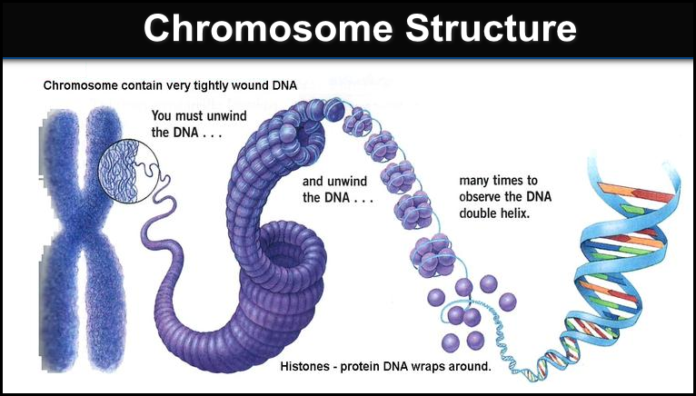
Chromosomes are thread-like structures found in the nucleus of eukaryotic cells. They are composed of DNA molecules tightly coiled and condensed around histone proteins. Each human cell typically contains 46 chromosomes, which are organized into 23 pairs (one set inherited from each parent).
Chromosome Structure
Each chromosome is a highly organized and intricate structure, consisting of several key components that enable its proper functioning during cell division and maintenance of genetic integrity.
The major components of a chromosome are:
a. Chromatids
A chromosome is made up of two identical chromatids, which are tightly coiled and condensed structures composed of DNA and proteins. These chromatids are joined together at a region called the centromere. During cell division, the chromatids separate and become individual chromosomes in the daughter cells, ensuring that each new cell receives a complete set of genetic information.
b. Centromere
The centromere is a constricted region that serves as a crucial attachment point for the spindle fibers during cell division. These spindle fibers are microtubule structures that play a vital role in the segregation of chromosomes during mitosis and meiosis. The centromere acts as a kinetochore, providing a platform for the spindle fibers to bind and exert the necessary forces to pull the chromatids apart and distribute them equally to the daughter cells.
c. Telomeres
Telomeres are repetitive DNA sequences found at the extreme ends of chromosomes. They consist of multiple repeats of a specific DNA sequence (TTAGGG in humans) and are associated with proteins called shelterins.
Telomeres serve two crucial functions:
Protection:
- They protect the chromosomes from degradation and fusion with other chromosomes by forming a cap-like structure at the ends.
- This cap prevents the cell’s DNA repair machinery from recognizing the chromosome ends as breaks that need to be repaired or fused.
Replication and Aging:
- Telomeres play a vital role in cell aging and replication.
- During each cell division, a small portion of the telomere is lost due to the inability of DNA polymerases to fully replicate the ends of linear DNA molecules.
- This progressive shortening of telomeres with each cell division acts as a biological clock, ultimately leading to cellular senescence (aging) or apoptosis (programmed cell death) when the telomeres become critically short.
The centromere and telomeres are essential structural components that ensure the proper segregation of chromosomes during cell division and maintain the integrity and stability of the genetic material over successive cell generations.
Types of Chromosomes
Chromosomes can be classified based on various criteria, including:
- Autosomal vs. Sex Chromosomes
- Autosomal chromosomes: Present in both males and females
- Sex chromosomes: Determine the sex of an organism (e.g., X and Y in humans)
- Morphology
- Metacentric: Centromere in the middle
- Submetacentric: Centromere slightly off-center
- Acrocentric: Centromere near one end
- Telocentric: Centromere at the end
- Special Types
- B chromosomes: Extra chromosomes found in some species
- Polytene chromosomes: Giant chromosomes found in certain tissues
- Lampbrush chromosomes: Large chromosomes found in oocytes
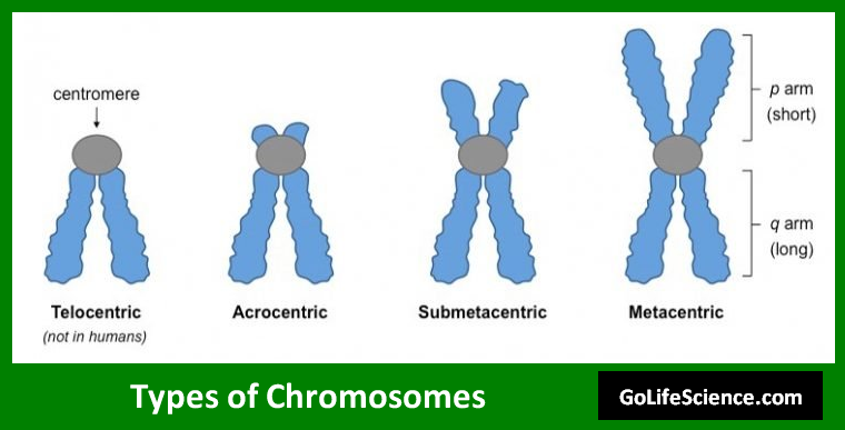
| Type | Location of Centromere | Arm Ratio | Example |
|---|---|---|---|
| Metacentric | Middle | 1:1 | Human chromosome 1 |
| Submetacentric | Off-center | 1:1.5 to 1:3 | Human chromosome 5 |
| Acrocentric | Near one end | 3:1 | Human chromosome 13 |
| Telocentric | At the end, | Essentially single-armed | Found in some insects and plants |
Chromosome Numbers
The number of chromosomes varies widely among different species. This number, known as the chromosome count or chromosome number, is typically represented as 2n for diploid organisms, where n is the number of unique chromosomes.
a. Notable Chromosome Numbers
- Humans: 2n = 46 (23 pairs)
- Chimpanzees: 2n = 48
- Domestic cat: 2n = 38
- Fruit fly (Drosophila melanogaster): 2n = 8
- Adder-tongue fern: 2n = 1260 (highest known chromosome count)
b. Ploidy Levels
Organisms can have different ploidy levels, referring to the number of complete sets of chromosomes in their cells:
- Haploid (n): One set of chromosomes (e.g., gametes)
- Diploid (2n): Two sets of chromosomes (most animals and plants)
- Polyploid (>2n): More than two sets of chromosomes (common in plants)
The number of chromosomes contributes to the determination of the taxonomic position of a plant or animal species. The chromosome number differs from species and it may range from two to several hundred.
The smallest number of chromosomes is found in Ascaris megalocephaly, having two somatic chromosomes i.e. one haploid chromosome.
The maximum number of chromosomes is found in protozoan. The group aggregate possesses more than 300 chromosomes while in Radiolaria, the number even reaches up to 1600.
In Animals
| Organism | Species | Diploid (2N) Chromosome Number |
| Man | Homo sapiens | 46 |
| Cat | Felix domesticus | 38 |
| Cattle | Bos taurus | 60 |
| Chicken | Gallus domesticus | 78 |
| Dog | Canis familiaris | 78 |
| Donkey | Equus asinus | 62 |
| Frog | Rana pipiens | 26 |
| Fruit fly | Drosophila melanogaster | 8 |
| Grasshopper | Melanoplus differentialis | 24 |
| Honeybee | Apis mellifera | 32 |
| Horse | Equus calibus | 64 |
| House fly | Musca domestica | 12 |
| Mosquito | Culex pipiens | 6 |
| Rabbit | Oryctolagus cuniculus | 44 |
| Red ant | Formica sanguinea | 48 |
| Rhesus monkey | Macaca mulatta | 42 |
In Plants:
| Organism | Species | Diploid (2N) Chromosome number |
| Alfalfa | Medicago sativa | 32 |
| Avocado | Persea americana | 24 |
| Barley | Hordeum vulgare | 14 |
| Bermudagrass | Cynodon dactylon | 36 |
| Broad bean | Vicia faba | 12 |
| Cashew | Anacardium occidentale | 42 |
| Corn (maize) | Zea mays | 20 |
| Cotton, American-Egyptian | Gossypium barbadense | 52 |
| Cotton, upland | Gossypium hirsutum | 52 |
| Durian | Durio zibethinus | 56 |
| Flax | Linum usitatissimum | 30 |
| Garden pea | Pisum sativum | 14 |
| Grape | Vitis vinifera | 38 |
| Guava | Psidium guajava | 22 |
| Kidney bean | Phaseolus vulgaris | 22 |
| Mango | Mangifera indica | 40 |
| Oats, white | Avena sativa | 42 |
| Oats, red | Avena byzantina | 42 |
| Onion | Allium cepa | 16 |
| Papaya | Carica papaya | 18 |
| Peanut | Arachis hypogaea | 40 |
| Pineapple | Ananas comosus | 50 |
| Potato | Solanum tuberosum | 48 |
| Rice | Oryza sativa | 24 |
| Rye | Secale cereale | 14 |
| Sorghum | Sorghum vulgare | 20 |
| Soybean | Glycine max | 40 |
| Squash | Cucurbita pepo | 40 |
| Sugar beet | Beta vulgaris | 18 |
| Sugar cane | Saccharum officinarum | 80 |
| Tamarind | Tamarindus indica | 24 |
| Tobacco | Nicotiana tabacum | 48 |
| Tomato | Lycopersicon esculentum | 24 |
| Wheat, durum | Triticum durum | 28 |
| Wheat, common | Triticum vulgare | 42 |
| Wheat, club | Triticum compactum | 42 |
Chemical Composition
- The chromosomes are mainly composed of nucleic acids and proteins.
- The DNA and histidine combine together and form deoxyribonucleoprotein, which is 90% of the chromosome, with DNA being 35% and histone protein being about 55%.
- The remaining 10% of a chromosome is termed as a residual chromosome.
- Stedman (1934) reported another type of protein called Chromosomin in addition to histone or protamines.
Chromosomal Abnormalities
Chromosomal abnormalities can occur due to errors in cell division or environmental factors. These abnormalities can be numerical (changes in chromosome number) or structural (changes in chromosome structure).
a. Numerical Abnormalities
- Aneuploidy: The presence of an abnormal number of chromosomes
- Monosomy: Missing one chromosome (2n-1)
- Trisomy: Extra chromosome (2n+1)
- Polyploidy: More than two complete sets of chromosomes
- Triploidy (3n)
- Tetraploidy (4n)
b. Structural Abnormalities
- Deletions: Loss of a chromosome segment
- Duplications: Extra copies of a chromosome segment
- Inversions: Reversal of a chromosome segment
- Translocations: Exchange of segments between non-homologous chromosomes
c. Chromosomal Abnormality Rate Formula
The rate of chromosomal abnormalities in a population can be calculated using the following formula:
Abnormality Rate = (Number of individuals with abnormalities / Total population size) × 100
This rate is often expressed as a percentage and can vary significantly depending on the specific abnormality and population studied.
Role of Chromosomes in Cell Division
Chromosomes play a crucial role in cell division, ensuring the accurate distribution of genetic material to daughter cells. The two main types of cell division are mitosis and meiosis.
a. Mitosis
Mitosis is the process of cell division that results in two genetically identical daughter cells. The stages of mitosis are:
- Prophase
- Metaphase
- Anaphase
- Telophase
During mitosis, chromosomes condense, align at the cell’s equator, and then separate, with one copy of each chromosome moving to opposite poles of the cell.
b. Meiosis
Meiosis is a specialized type of cell division that produces gametes (reproductive cells) with half the number of chromosomes as the parent cell. Meiosis involves two rounds of division:
- Meiosis I
- Meiosis II
Meiosis is crucial for sexual reproduction and genetic diversity as it involves the process of crossing over, where genetic material is exchanged between homologous chromosomes.
c. Chromosome Segregation Accuracy
The accuracy of chromosome segregation during cell division can be expressed using the following formula:
Segregation Accuracy = (Number of cells with correct chromosome number / Total number of cells) × 100
High segregation accuracy is essential for maintaining genomic stability and preventing chromosomal abnormalities.
Chromosomes and Heredity
Chromosomes are the physical basis of heredity, carrying the genes that determine an organism’s traits. The principles of inheritance, first described by Gregor Mendel, are directly related to the behavior of chromosomes during meiosis and fertilization.
a. Key Concepts in Chromosomal Inheritance
- Alleles: Alternative forms of a gene
- Homologous chromosomes: Pairs of chromosomes with the same genes in the same order
- Dominant and recessive traits: Determined by the interaction of alleles
- Linkage: The tendency of genes on the same chromosome to be inherited together
b. Punnett Square
A Punnett square is a tool used to predict the possible genotypes and phenotypes of offspring based on the genotypes of the parents. For example, consider a cross between two heterozygous individuals (Aa × Aa):
| A | a | |
| A | AA | Aa |
| a | Aa | aa |
This Punnett square shows the possible genotypes of offspring: 25% AA, 50% Aa, and 25% Aa.
Chromosome Mapping and Sequencing
Chromosome mapping and sequencing are crucial techniques in modern genetics, allowing researchers to identify the location and sequence of genes on chromosomes.
a. Chromosome Mapping Techniques
- Linkage mapping: Uses genetic recombination frequencies to determine the relative positions of genes
- Physical mapping: Directly localizes DNA sequences on chromosomes using techniques like fluorescence in situ hybridization (FISH)
- Radiation hybrid mapping: Uses radiation-induced chromosome breakage to map genes
b. Chromosome Sequencing
The sequencing of entire chromosomes has revolutionized our understanding of genetics and genomics. The Human Genome Project, completed in 2003, sequenced all human chromosomes, providing a wealth of information about our genetic makeup.
c. Genetic Distance Formula
Genetic distance, used in linkage mapping, can be calculated using the following formula:
Genetic Distance (in centiMorgans) = (Number of recombinant offspring / Total number of offspring) × 100
This formula helps geneticists determine the relative positions of genes on a chromosome.
Packaging of DNA
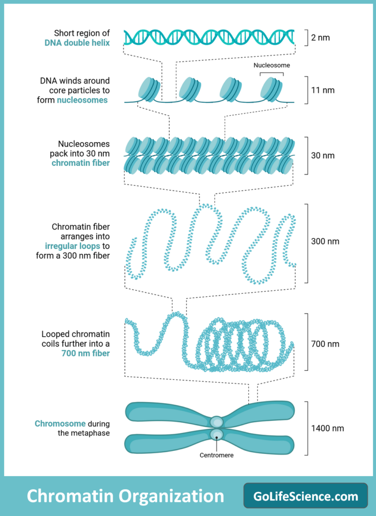
DNA molecules are incredibly long and must be packaged efficiently to fit within the confines of the cell nucleus. This packaging process involves several levels of organization, from the basic DNA double helix to the highly condensed chromosomal structure.
a. DNA Double Helix
The DNA molecule is a double-stranded helix composed of two complementary strands of nucleotides (adenine, guanine, cytosine, and thymine). The two strands are held together by hydrogen bonds between the complementary base pairs (A-T and G-C).
b. Nucleosomes and Chromatin
The first level of DNA packaging involves the wrapping of DNA around histone proteins to form nucleosomes. Histones are small, positively charged proteins that act as spools for the DNA to coil around. A DNA segment (roughly 146 base pairs) is encircled by a histone octamer, which is made up of two copies of each of the four histone proteins (H2A, H2B, H3, and H4) to form each nucleosome.
Chromatin is the term for the structure formed by linker DNA segments connecting nucleosomes. Chromatin can exist in two states:
- Euchromatin: This is a loosely packed form of chromatin that is accessible to transcription factors and other regulatory proteins, allowing for gene expression.
- Heterochromatin: This is a highly condensed and tightly packed form of chromatin, which is generally inaccessible for gene expression. Heterochromatin is found in regions of the genome that are transcriptionally inactive or contain repetitive DNA sequences.
c. Higher-Order Packaging
The packaging of DNA continues at higher levels of organization. Chromatin fibers are further folded and coiled into loops and domains, creating a more compact structure. These coiled structures are then further condensed and compacted into the characteristic chromosome shape observed during cell division.
Evolutionary Aspects of Chromosomes
Chromosomes play a significant role in evolution, with changes in chromosome structure and number contributing to speciation and adaptation.
a. Chromosomal Evolution Mechanisms
- Chromosomal rearrangements: Inversions, translocations, and fusions can lead to reproductive isolation
- Changes in chromosome number: Polyploidy events can result in new species, especially in plants
- Centromere repositioning: This can occur without changing gene order but may affect chromosome segregation
b. Karyotype Evolution
The karyotype, or the complete set of chromosomes in an organism, can evolve over time. Comparative genomics allows researchers to trace the evolutionary history of chromosomes across species.
c. Chromosomal Speciation Rate
The rate of speciation due to chromosomal changes can be estimated using the following formula:
Chromosomal Speciation Rate = (Number of new species arising from chromosomal changes / Total number of speciation events) × 100
Population size and environmental conditions are two factors that affect this rate, which varies greatly among different taxonomic groups.
Chromosomes in Medicine and Research
Chromosomes are of great importance in medical diagnostics and research, providing insights into genetic disorders, cancer, and other diseases.
a. Chromosomal Disorders
Many genetic disorders are caused by chromosomal abnormalities, including:
- Down syndrome (Trisomy 21)
- Turner syndrome (Monosomy X)
- Klinefelter syndrome (XXY)
- Cri-du-chat syndrome (Deletion on chromosome 5)
b. Karyotyping
Karyotyping is a technique used to visualize and analyze an individual’s chromosomes. It is used in prenatal testing, cancer diagnosis, and genetic counseling.
c. Chromosomes in Cancer Research
Cancer often involves chromosomal abnormalities, such as:
- Philadelphia chromosome in chronic myeloid leukemia
- HER2 amplification in breast cancer
- p53 deletions in various cancers
d. Chromosomal Instability Index
In cancer research, chromosomal instability can be quantified using the Chromosomal Instability Index (CIN):
CIN = (Number of cells with abnormal chromosome number / Total number of cells analyzed) × 100
A higher CIN value indicates greater genomic instability, which is often associated with more aggressive cancers.
Functions of Chromosomes
What is the function of a chromosome in a cell? The chromosome functions are given below
- They control the physiological behavior of an organism with the help of genes present in them.
- We know each chromosome is made up of DNA and this DNA, by replication, gives rise to messenger RNA (mRNA), which carries the genetic information in the form of code.
- This mRNA comes out of the nuclear wall and into the cytoplasm, where it helps form a particular kind of protein needed by the cell or body.
- Sometimes regions within the chromosomes change their position, which leads to genetic effects or mutations. Such an effect is termed a position effect, which is due to shifting in the position of the heterochromatin and euchromatin parts.
- Heterochromatin aids in the formation of the nucleus.
- They are a hereditary vehicle, carrying genetic information from one generation to the next.
Frequently Asked Questions (FAQs)
What are chromosomes?
Chromosomes are long, thread-like structures made of DNA and proteins. They contain the genetic information necessary for the growth, development, and reproduction of an organism.
How many chromosomes do humans have?
Humans typically have 46 chromosomes, arranged in 23 pairs. Each parent contributes one chromosome to each pair.
What is the function of chromosomes?
Chromosomes carry genes, the units of heredity. They ensure DNA is accurately copied and distributed during cell division, which is crucial for growth, development, and reproduction.
What are the types of chromosomes?
There are two types of chromosomes: autosomes and sex chromosomes. Autosomes are non-sex chromosomes, while sex chromosomes (X and Y) determine an individual’s sex.
What happens if there is an abnormal number of chromosomes?
An abnormal number of chromosomes can lead to genetic disorders. For example, Down syndrome is caused by an extra copy of chromosome 21, resulting in 47 chromosomes instead of the usual 46.
Final words about Chromosomes
Chromosomes are the cornerstone of genetics and heredity, playing a crucial role in the storage, transmission, and expression of genetic information. From their intricate structure to their complex behavior during cell division, chromosomes continue to fascinate scientists and drive advances in fields such as medicine, agriculture, and evolutionary biology.
As our understanding of chromosomes deepens, we unlock new possibilities for treating genetic disorders, developing targeted cancer therapies, and unraveling the mysteries of life itself. The study of chromosomes remains a vibrant and essential area of research, promising continued discoveries and innovations that will shape the future of biology and medicine.
By exploring the various aspects of chromosomes, from their basic structure to their role in evolution and disease, we gain a greater appreciation for the complexity and elegance of life at the molecular level. As we continue to unravel the secrets hidden within our chromosomes, we move closer to a more complete understanding of ourselves and the living world around us.

