Muscles are essential for movement, posture, and stability in the human body. They allow us to walk, run, lift, and perform a wide range of activities. Without muscle, the human body would be unable to function properly. This note describes the structure of muscles, as well as their types, contractions, and functions.
But what exactly is muscle, and how does it work? In this post, we’ll delve into the structure and function of muscle tissue, including the different types of muscle and how they contribute to the overall functioning of the body. We’ll also explore the process of muscle contraction and the role it plays in movement.
By the end of this post, you’ll have a better understanding of the complex and vital role that muscle plays in the human body.
Structure of Muscle cell
Muscle cells have a complicated and well-organized structure, with each part doing a certain job.

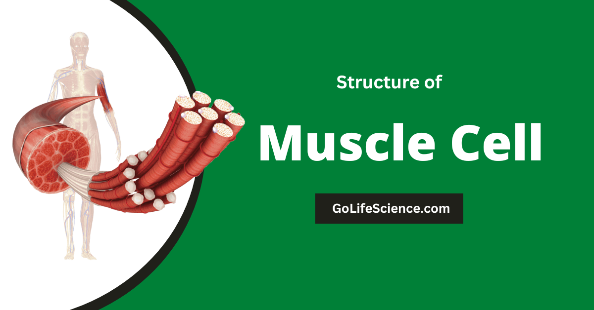
- Fascicle structure: Muscles are made up of long, cylindrical bundles of muscle fibers called fascicles. These fascicles are surrounded by connective tissue called the epimysium. These fascicles give muscles their shape and allow for the coordination of muscle contractions.
- Muscle fibers: Each fascicle is made up of individual muscle fibers, which are long, cylindrical cells. These muscle fibers are surrounded by a layer of connective tissue called the perimysium.
- Myofibrils: Inside each muscle fiber, there are smaller structures called myofibrils. These myofibrils are made up of protein filaments called actin and myosin. Myofibrils are smaller structures within muscle fibers that are made up of protein filaments called actin and myosin. These filaments are responsible for muscle contraction.
- Sarcomeres: The myofibrils are further organized into repeating units called sarcomeres. Sarcomeres are the basic functional unit of muscle and are found within the myofibrils. They contain the actin and myosin filaments and are responsible for the contraction and relaxation of muscle.
- Filaments: Actin and myosin are protein filaments that are found within the sarcomeres of muscle. When a muscle contracts, the actin and myosin filaments slide past each other, resulting in the shortening of the muscle.
Understanding the structure of a muscle is crucial for understanding how a muscle cell functions and is able to produce movement. In the next section, we’ll explore the process of muscle contraction in more detail.
Types of Muscle
There are three main types of muscular tissue in the human body: skeletal, smooth, and cardiac. Skeletal muscle is the type of muscle we most commonly think of when we think of muscles. It is responsible for movement, posture, and stability.

a. Skeletal Muscle
Skeletal muscle is attached to the bones of the skeleton and is responsible for movement of the limbs and other parts of the body. The skeletal muscle structure is very simple.
- Skeletal muscle is the type of muscle that is most commonly thought of when we think of muscle.
- It is responsible for movement, posture, and stability in the human body.
- Skeletal muscle is attached to the bones of the skeleton and is responsible for movement of the limbs and other parts of the body.
- Skeletal muscle is made up of long, cylindrical muscle fibers that are surrounded by connective tissue called the perimysium.
- Each muscle fiber is made up of smaller structures called myofibrils, which are organized into repeating units called sarcomeres.
- The sarcomeres contain protein filaments called actin and myosin, which are responsible for muscle contraction.
- Skeletal muscle is under conscious control and can be voluntarily contracted or relaxed.
- Skeletal muscle is highly adaptable and can change in size and strength in response to different types of physical activity.
- Maintaining healthy skeletal muscle tissue is important for overall health and well-being. This can be achieved through regular physical activity and a balanced diet.
b. Smooth Muscle
Smooth muscle is found in the walls of organs and structures such as the digestive tract, blood vessels, and the uterus. It functions to move substances through the body and regulate the size of openings in the body.
- Smooth muscle is a type of involuntary muscle that is found in the walls of organs and structures such as the digestive tract, blood vessels, and the uterus.
- It functions to move substances through the body and regulate the size of openings in the body.
- Smooth muscle is not under conscious control and is stimulated by the autonomic nervous system.
- Smooth muscle is made up of small, spindle-shaped cells that are arranged in sheets or layers.
- Like skeletal muscle, smooth muscle contains myofibrils and sarcomeres, but the arrangement of these structures is different.
- The contraction of smooth muscle is slower and more sustained than that of skeletal muscle.
- Smooth muscle is important for maintaining the proper functioning of various organs and structures in the body.
- Maintaining healthy smooth muscle tissue is important for overall health and well-being. This can be achieved through a healthy lifestyle and avoiding behaviors that may damage smooth muscle tissue, such as smoking.
c. Cardiac muscle
Cardiac muscle is found in the heart and is responsible for pumping blood throughout the body. Cardiac muscle is unique in that it is able to contract without being consciously controlled.
- Cardiac muscle is a type of involuntary muscle that is found in the heart.
- It is responsible for pumping blood throughout the body.
- Cardiac muscle is unique in that it is able to contract without being consciously controlled.
- It is stimulated by the electrical activity of the heart, which is regulated by the sinoatrial (SA) and atrioventricular (AV) nodes.
- Cardiac muscle cells are longer and more cylindrical than smooth muscle cells, and they are arranged in a branching pattern.
- Like skeletal and smooth muscle, cardiac muscle contains myofibrils and sarcomeres.
- The contraction of cardiac muscle is slower and more sustained than that of skeletal muscle.
- Maintaining healthy cardiac muscle tissue is essential for overall health and well-being. This can be achieved through a healthy lifestyle, including regular physical activity and a balanced diet, and managing stress.
- Cardiac muscle can be damaged by various factors, including high blood pressure, high cholesterol, and smoking. It is important to address these risk factors to maintain a healthy heart.
Each type of muscle tissue has its own unique characteristics and functions, but they all play a crucial role in the overall functioning of the body. In the next section, we’ll take a closer look at the structure of a muscle.
Contraction of a muscle
Muscle contraction is the process by which a muscle produces movement. It is a complex process that involves a series of steps at the molecular level.
a. Excitation-contraction coupling
Excitation-contraction coupling is the process by which the nervous system tells a muscle cell to get ready to contract and starts the process.

This process involves several steps:
- Nerve impulse: The process of excitation-contraction coupling begins with the activation of a muscle cell by a nerve impulse. This impulse is carried by a neuron, or nerve cell, and is transmitted to the muscle cell through a structure called the neuromuscular junction.
- Action potential: When the nerve impulse reaches the muscle cell, it initiates an action potential. An action potential is a brief, rapid change in the electrical charge of the muscle cell membrane.
- Calcium release: The action potential triggers the release of calcium ions from storage structures called the sarcoplasmic reticulum. These calcium ions bind to proteins called troponin, which are found on the actin filaments.
- Myosin-actin interaction: The binding of calcium ions to troponin causes a conformational change in the troponin-tropomyosin complex, exposing the binding sites on the actin filaments. This allows the myosin filaments to bind to the actin filaments and initiate muscle contraction.
Overall, excitation-contraction coupling is a complicated process that is important for movement and muscle contraction.
b. Sliding filament theory
The sliding filament theory is a model that explains how molecular-level muscle contraction works. This theory says that the actin and myosin filaments in the sarcomeres contract the muscle when they slide past each other.

This process involves several steps:
- Cross-bridge formation: When a muscle is stimulated to contract, the myosin filaments form “cross-bridges” with the actin filaments. These cross-bridges are formed by the “heads” of the myosin filaments, which bind to the actin filaments.
- ATP hydrolysis: The formation of cross-bridges requires the hydrolysis (breakdown) of ATP, a molecule that stores energy in cells. When ATP is hydrolyzed, it releases energy that is used to power the movement of the myosin filaments.
- Power stroke: Once the cross-bridges are formed and ATP is hydrolyzed, the myosin filaments undergo a movement called the power stroke. During this movement, the myosin heads bend and pull the actin filaments toward the center of the sarcomere. This results in the shortening of the muscle.
- Cross-bridge detachment: After the power stroke is completed, the myosin heads detach from the actin filaments and return to their original position. This allows the muscle to relax and return to its resting state.
Overall, the sliding filament theory explains how muscle contraction occurs through the movement of the actin and myosin filaments within the sarcomeres. This process is needed for muscles to move, and it is powered by the breakdown of ATP by water.
Basically, muscle contraction is a complex process that is essential for movement and the functioning of the human body.
Conclusion
In conclusion, muscle is a vital tissue in the human body that is responsible for movement, posture, and stability. There are three main types of muscle tissue: skeletal, smooth, and cardiac, each with its own unique characteristics and functions.
Muscles have a complicated and well-organized structure, with each part doing a certain job. When a muscle contracts, it moves. This is caused by a process called excitation-contraction coupling, which is set off by the nervous system.
The actual process of muscle contraction occurs according to the sliding filament theory, where the actin and myosin filaments within the sarcomeres slide past each other.
Maintaining healthy muscle tissue is important for overall health and well-being. This includes engaging in regular physical activity, eating a balanced diet, and managing stress.
By better understanding the structure of muscles and their functions, we can better understand how to take care of our muscles and keep them functioning properly.
