Nucleic Acid Structures

Nucleic acids are the fundamental molecules of life, responsible for storing and transmitting genetic information. They are composed of two main types: deoxyribonucleic acid (DNA) and ribonucleic acid (RNA). These molecules are essential for the development, functioning, and reproduction of all known living organisms. Understanding the structure of nucleic acids is crucial for comprehending how genetic information is encoded, replicated, and expressed. This article delves into the intricate world of nucleic acid structures, exploring their composition, organization, and the significance of their three-dimensional configurations.

The Building Blocks of Nucleic Acids

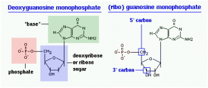
Nucleic acids are polymers made up of repeating units called nucleotides. Each nucleotide consists of three components:

Nucleic acids Structures
  1. Phosphate Group: A phosphorus atom bonded to four oxygen atoms, providing a negative charge.
  2. Sugar Molecule: A five-carbon sugar, either ribose (in RNA) or deoxyribose (in DNA).
  3. Nitrogenous Base: A nitrogen-containing base that can be either a purine (adenine or guanine) or a pyrimidine (cytosine, thymine in DNA, or uracil in RNA).

The combination of a sugar molecule and a nitrogenous base is called a nucleoside. When a phosphate group is attached to the nucleoside, it forms a nucleotide.

The basic structure of nucleic acids is Nitrogenous bases, the sugar moiety, and the Phosphate molecule.

  • Nucleoside = Nitrogen base + Sugar
  • Nucleotide = Nucleoside (Nitrogen base + Sugar) + Phosphate molecule

Nucleotides are the building blocks of all nucleic acids. Nucleotides have a distinctive structure composed of three components covalently bound together:

  • Nitrogenous bases– Pyrimidine (one ring)  and Purine (two rings)
  • Sugar moiety – Ribose or Deoxyribose
  • Phosphate molecule

The Phosphodiester Bond

Nucleotides are linked together through phosphodiester bonds, which form between the phosphate group of one nucleotide and the hydroxyl group of the sugar molecule of another nucleotide. This linkage creates a sugar-phosphate backbone, with the nitrogenous bases extending from it. The sequence of these bases along the backbone encodes genetic information.

Structure of Nucleosides and Nucleotides

Nucleosides

A nucleoside is composed of two main components:

  1. Sugar Molecule: The sugar in a nucleoside is a pentose sugar, which can be either ribose (in RNA) or deoxyribose (in DNA). Ribose has a hydroxyl group (-OH) at the 2′ position, while deoxyribose has a hydrogen atom (-H) at the same position.
  2. Nitrogenous Base: The nitrogenous base is a nitrogen-containing molecule that can be either a purine or a pyrimidine. The purines include adenine (A) and guanine (G), while the pyrimidines include cytosine (C), thymine (T) in DNA, and uracil (U) in RNA.

The bond between the sugar molecule and the nitrogenous base is called a glycosidic bond. This bond forms between the 1′ carbon of the sugar and the nitrogen atom (N1 for pyrimidines and N9 for purines) of the base.

Nucleotides

A nucleotide is a nucleoside with one or more phosphate groups attached to the sugar molecule. The phosphate group is typically attached to the 5′ carbon of the sugar, forming a phosphoester bond. Nucleotides can have one, two, or three phosphate groups, referred to as nucleoside monophosphate (NMP), nucleoside diphosphate (NDP), and nucleoside triphosphate (NTP), respectively.

The general structure of a nucleotide can be summarized as:

  • Sugar (Ribose or Deoxyribose)
  • Nitrogenous Base (Purine or Pyrimidine)
  • Phosphate Group(s)

Types of Nucleosides and Nucleotides

Nucleosides

  1. Adenosine: Composed of adenine and ribose.
  2. Guanosine: Composed of guanine and ribose.
  3. Cytidine: Composed of cytosine and ribose.
  4. Uridine: Composed of uracil and ribose.
  5. Thymidine: Composed of thymine and deoxyribose (found in DNA).

Nucleotides

  1. Adenosine Monophosphate (AMP): Adenosine with one phosphate group.
  2. Adenosine Diphosphate (ADP): Adenosine with two phosphate groups.
  3. Adenosine Triphosphate (ATP): Adenosine with three phosphate groups.
  4. Guanosine Monophosphate (GMP): Guanosine with one phosphate group.
  5. Cytidine Monophosphate (CMP): Cytidine with one phosphate group.
  6. Uridine Monophosphate (UMP): Uridine with one phosphate group.
  7. Thymidine Monophosphate (TMP): Thymidine with one phosphate group.

The Double Helix: DNA’s Iconic Structure

Discovery of the Double Helix

The double helix structure of DNA was first proposed by James Watson and Francis Crick in 1953, based on X-ray diffraction images produced by Rosalind Franklin and Maurice Wilkins. This model revolutionized the field of molecular biology, providing a clear understanding of how genetic information is stored and replicated.

Key Features of the DNA Double Helix

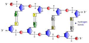
  1. Antiparallel Strands: The two strands of DNA run in opposite directions, with one strand running 5′ to 3′ and the other running 3′ to 5′. This antiparallel arrangement is crucial for DNA replication and transcription.
  2. Complementary Base Pairing: The nitrogenous bases on one strand pair with specific bases on the opposite strand through hydrogen bonds. Adenine pairs with thymine (A-T), and cytosine pairs with guanine (C-G). This complementary base pairing ensures the accurate replication of DNA.
  3. Major and Minor Grooves: The double helix has two grooves of different widths, known as the major and minor grooves. These grooves provide binding sites for proteins that regulate gene expression and DNA replication.

DNA Conformations

DNA can exist in several different conformations, depending on environmental conditions and sequence:

  1. B-DNA: The most common and stable form of DNA, characterized by a right-handed helix with 10 base pairs per turn.
  2. A-DNA: A shorter, wider right-handed helix that forms under dehydrating conditions.
  3. Z-DNA: A left-handed helix that can form in sequences with alternating purines and pyrimidines, such as GCGCGC.

RNA Structures: Versatility in Function

Single-Stranded Nature of RNA

Unlike DNA, RNA is typically single-stranded. However, it can fold back on itself to form complex secondary and tertiary structures, which are essential for its function. The single-stranded nature of RNA allows it to adopt a variety of shapes, enabling it to perform diverse roles in the cell.

Types of RNA and Their Structures

  1. Messenger RNA (mRNA): mRNA carries genetic information from DNA to the ribosome, where it is translated into protein. It is generally linear but can form secondary structures that regulate its stability and translation efficiency.
  2. Transfer RNA (tRNA): tRNA is responsible for bringing amino acids to the ribosome during protein synthesis. It has a cloverleaf secondary structure that folds into an L-shaped tertiary structure, with one end binding to the amino acid and the other end recognizing the codon on the mRNA.
  3. Ribosomal RNA (rRNA): rRNA is a major component of ribosomes, the cellular machinery for protein synthesis. It forms complex secondary and tertiary structures that are crucial for ribosome assembly and function.
  4. Small Nuclear RNA (snRNA): snRNA is involved in the splicing of pre-mRNA in the nucleus. It forms part of the spliceosome, a complex that removes introns from pre-mRNA.
  5. MicroRNA (miRNA): miRNA regulates gene expression by binding to complementary sequences on mRNA, leading to its degradation or inhibition of translation. It forms hairpin structures that are processed into mature miRNA.

RNA Folding and Stability

RNA folding is driven by base pairing and base stacking interactions. The formation of secondary structures, such as hairpins, loops, and bulges, is stabilized by hydrogen bonds and van der Waals forces. Tertiary structures are stabilized by additional interactions, including metal ion binding and long-range base pairing.

Nucleic Acid-Protein Interactions

DNA-Protein Interactions

Proteins that interact with DNA play crucial roles in gene regulation, DNA replication, and repair. These interactions are often mediated by specific DNA-binding domains, such as:

  1. Helix-Turn-Helix: Found in many transcription factors, this domain consists of two α-helices connected by a short turn. One helix fits into the major groove of DNA, while the other stabilizes the interaction.
  2. Zinc Finger: A small protein domain stabilized by a zinc ion, which interacts with specific DNA sequences.
  3. Leucine Zipper: A dimerization domain that allows two protein subunits to bind to adjacent major grooves of DNA.

RNA-Protein Interactions

RNA-binding proteins are essential for RNA processing, transport, and function. These proteins recognize specific RNA sequences or structures, often through RNA-binding domains such as:

  1. RNA Recognition Motif (RRM): A common domain that binds single-stranded RNA.
  2. Double-Stranded RNA-Binding Domain (dsRBD): Binds to double-stranded RNA, often found in proteins involved in RNA interference.
  3. KH Domain: Binds to single-stranded RNA and is involved in RNA splicing and translation regulation.

Nucleic Acid Structures in Disease

Mutations and Genetic Disorders

Mutations in DNA sequences can lead to changes in protein structure and function, resulting in genetic disorders. For example:

  1. Point Mutations: A single nucleotide change can lead to amino acid substitutions, as seen in sickle cell anemia.
  2. Insertions and Deletions: These can cause frameshift mutations, leading to nonfunctional proteins, as in cystic fibrosis.
  3. Chromosomal Rearrangements: Large-scale changes in chromosome structure can lead to diseases like chronic myeloid leukemia.

RNA in Disease

RNA molecules can also be involved in disease processes. For example:

  1. MicroRNA Dysregulation: Abnormal expression of miRNAs has been linked to cancer, cardiovascular diseases, and neurological disorders.
  2. RNA Viruses: Viruses like HIV and SARS-CoV-2 use RNA as their genetic material, and understanding their RNA structures is crucial for developing antiviral therapies.

Techniques for Studying Nucleic Acid Structures

  • X-ray Crystallography: X-ray crystallography is a powerful technique for determining the atomic structure of nucleic acids. It involves crystallizing the nucleic acid and exposing it to X-rays, which diffract to produce a pattern that can be used to reconstruct the 3D structure.
  • Nuclear Magnetic Resonance (NMR) Spectroscopy: NMR spectroscopy is used to study the structure and dynamics of nucleic acids in solution. It provides information on the distances between atoms and the overall shape of the molecule.
  • Cryo-Electron Microscopy (Cryo-EM): Cryo-EM is a technique that allows for the visualization of large nucleic acid-protein complexes at near-atomic resolution. It involves freezing the sample and imaging it with an electron microscope.
  • Computational Modeling: Computational methods, such as molecular dynamics simulations and bioinformatics, are used to predict and analyze nucleic acid structures. These methods complement experimental techniques and provide insights into the dynamic behavior of nucleic acids.

Applications of Nucleic Acid Structures in Biotechnology

  • Gene Editing: Understanding nucleic acid structures has enabled the development of gene-editing technologies, such as CRISPR-Cas9. This system uses a guide RNA to direct the Cas9 protein to specific DNA sequences, where it introduces double-strand breaks, allowing for precise gene editing.
  • Drug Design: Knowledge of nucleic acid structures is essential for designing drugs that target DNA or RNA. For example, anticancer drugs like cisplatin bind to DNA and disrupt its structure, preventing cancer cells from replicating.
  • Synthetic Biology: Synthetic biology involves designing and constructing new biological parts, devices, and systems. Understanding nucleic acid structures allows scientists to engineer synthetic genes, regulatory elements, and even entire genomes.

Conclusion

Nucleic acid structures are the foundation of life, encoding the genetic information that dictates the development, function, and reproduction of all living organisms. From the iconic double helix of DNA to the versatile folds of RNA, these molecules exhibit a remarkable diversity of forms and functions. Advances in structural biology have provided unprecedented insights into the molecular mechanisms underlying genetic information storage, replication, and expression. As our understanding of nucleic acid structures continues to grow, so too does our ability to harness this knowledge for applications in medicine, biotechnology, and synthetic biology. The study of nucleic acid structures is not only a fascinating scientific endeavor but also a key to unlocking the mysteries of life itself.

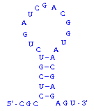
What about double-stranded RNA? RNAs are usually single-stranded, but many RNA molecules have a secondary structure in which intramolecular loops are formed by complementary base pairing. A simple example of this is shown in the figure to the right, and much more extensive and complex examples are known. Base pairing in RNA follows exactly the same principles as with DNA: the two regions involved in duplex formation are antiparallel to one another, and the base pairs that form are A-U and G-C.

image

OK, what about RNA-DNA hybrids? Can they form? The answer is yes. Complementary sequences of RNA and DNA readily anneal with one another to form duplexes. In fact, RNA-DNA hybrids are more stable than the corresponding DNA-DNA and RNA-RNA duplexes.

Similar Posts